Preliminary Research and Documentation
he following research and documentation is meant to get this project started. Please consider this a work in progress. Significant work is needed to move this project forward.
RequirementsComponents
StructureElectrodes (2) - Stainless steel or iron or aluminum electrodes.- DC Power Supply - A power supply unit that can provide a 5V DC power to the circuit.
- Multimeter - A multimeter to check voltage and
Materialampere o electricity. - Wires -
NaturalTocoralsconnectarethemadeelectrodes to the DC power supply. - Waste water - 1 liter of
calciumacarbonatehomogeneous(limestone).mixtureWeofneedwater,tooil,investigatevegetableifjuice,wesoil,can print using calcium carbonatemeat, andotheralcohol. - Gloves and protective eye wear.
Method
An electrocoagulation unit consists of an anode and a cathode that are are sturdy and eco-friendly. The structure must be sturdy enoughconnected to withstanda underwaterDC currentspower andsupply. impactWhen bycontaminated bigwater fish.flows Theinto coloringan mustelectrocoagulation look natural.
- From the
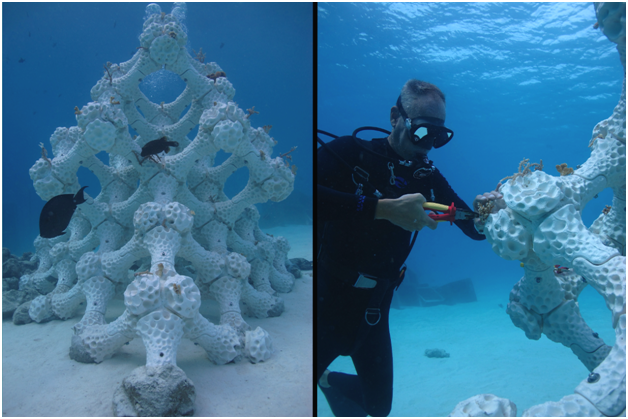
structure,anode,wemetallicneedionstoareensure that there is enough natural lighting in the crevices. Also we need to investigate if we can add artificial lighting and maybe even a camera to study polyps and other marine life. Rooting the structure- Natural corals tend to grow roots and growreleased into theincontaminated water.- On the
oceancathode,bed.waterTheis3Dhydrolyzed,printedformingcoralhydrogenmustgashave(H2)aandrootinghydroxylstructure(OH)thatparticles. - Electrons
bemovedrilled intofrom theoceancathodefloor.to the anode. Thiswilldestabilizesmakesurface charges on theoverallsuspendedstructuresolidssturdy enough to withstand under water currents and impact by big fish.(colloids).
Figure 31 - CoralWaste reefwater installationtreatment using electrocoagulation
When the electrons destabilize the surface charges on these suspended particles, the metal ions, along with the hydroxyl particles form complex compounds called flocs that includes metals and other contaminants. Colloids and emulsified oils combine with these flocs to form sludge. Depending on the chemical composition of the floc, the sludge can either rise to the top and float or sink to the bottom. Sludge can be removed physically from the sludge tanks and disposed off in an eco-friendly manner.
The water is now available for further filtering or reuse.
ReferencesFlocculation
InThe 1997process of water filtration also involves a process called flocculation. This process is usually done before the firstwater Sayais dereleased Malhainto expeditionthe waselectrocoagulation conductedunit. byFlocculants Wolfare Hilbertz,chemicals Thomasthat Goreau,are Kaiadded Hilbertzto contaminated water to form flocs and Carolinesludge.
Surprisingly these reefs were not dominated by any one group of corals, as is typical of most Indian Ocean reefs. Instead the coral populations consisted of small numbers of many different groups of corals, widely distributed. The larger corals were mostly rounded heads of Porites, or clumps of columnar Heliopora or Millepora, with smaller corals of many kinds around them. Many corals were observed to be loosely attached to the bottom, and many were being attacked by boring sponges, by several distinct coral diseases, or had algae overgrowing their edges.
Detailed information is available in the Saya de Malha Expedition (2002) report.
Also See
Dr. David Vaughan at the Mote Laboratory is growing coral 40 times faster than in the wild. The lab takes a coral that is about as big as a golf ball and cut it into 20 to 100 micro-fragments. Each fragment grows back in a few months. Detailed information and links are available in the Micro-Fragmenting report.


